Friday, 4 May 2018

Microbiology - bacterial Growth

Picture

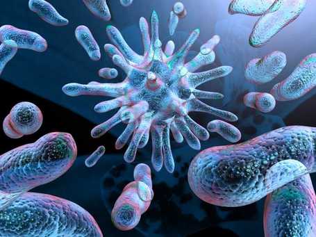
BACTERIAL GROWTH

Growth of Bacteria is the orderly increase of all the chemical constituents of the bacteria. Multiplication is the consequence of growth. Death of bacteria is the irreversible loss of ability to reproduce.

Bacteria are composed of proteins, carbohydrates, lipids, water and trace elements.
Factors Required for Bacterial Growth
The requirements for bacterial growth are:
(A)) Environmental factors affecting growth, and
(B) Sources of metabolic energy.

A. Environmental Factors affecting Growth:
1. Nutrients. Nutrients in growth media must contain all the elements necessary for the synthesis of new organisms. In general the following must be provided : (a) Hydrogen donors and acceptors, 

(b) Carbon source, (c) Nitrogen source, (d) Minerals : sulphur and phosphorus, (e) Growth factors: amino acids, purines, pyrimidines; vitamins, (f) Trace elements: Mg, Fe, Mn.

Growth Factors: 

A growth factor is an organic compound which a cell must contain in order to grow but which it is unable to synthesize. These substances are essential for the organism and are to be supplied as nutrients. Thiamine, nicotinic acid, folic acid and para-aminobenzoic acid are examples of growth factors.

Essential Metabolites: These metabolites are essential for growth of bacterium. These must be synthesized by the bacterium, or be provided in the medium. Mg, Fe and Mn are essential trace elements.

Autotrophs live only on inorganic substances, i.e. do not require organic nutrients for growth. They are not of medical importance.

Heterotrophs require organic materials for growth, e.g. proteins, carbohydrates, lipids as source of energy. All bacteria of medical importance belong to heterotrophs. Parasites may depend on the host for certain foods. Saprophytes grow, on dead organic matter.

2.   pH of the medium. Most pathogenic bacteria grow best in pH 7.2-7.4. Vibno cholerae can grow in pH 8.2-9.0.
Picture
3.   Gaseous Requirement
(a) Role of Oxygen. Bacteria may be classified into four groups on oxygen requirement :
(i)    Aerobes. They cannot grow without oxygen, e.g. Mycobacterium tuberculosis.
(ii)   Facultative anaerobes. These grow under both aerobic and anaerobic conditions. Most bacteria are facultative anaerobes, e.g. Enterobacteriaceae.
(iii)Anaerobes. They only grow in absence of free oxygen, e.g. Clostridium, Bacteroides.
(iv)   Microaerophils grow best in oxygen less than that present in the air, e.g. Campylobacter.

Aerobes and facultative anaerobes have the metabolic systems: (1) cytochrome systems for the metabolism of oxygen, (2) Superoxide dismutase, (3) catalase.

Anaerobic bacteria do not grow in the presence of oxygen. They do not use oxygen for growth and metabolism but obtain their energy from fermentation reactions. Anaerobic bacteria are killed by oxygen or toxic oxygen radicals. Multiple mechanisms play role for oxygen toxicity : (1) They do not have cytochrome systems for oxygen metabolism, (2) They may have low levels of superoxide dismutase, and (3) They may or may not have catalase.

(b) Carbon dioxide. All bacteria require CO2 for their growth. Most bacteria produce CO2. N. gonorrhoeae and N. meningitides and Br abortus grow better in presence of 5 per cent CO2.

4.   Temperature. Most bacteria are mesophilic. Mesophilic bacteria grow best at 30-37°C. Optimum temperature for growth of common pathogenic bacteria is 37°C. Bacteria of a species will not grow but may remain alive at a maximum and a minimum temperature.

5.   Ionic strength and osmotic pressure.

6.   Light. Optimum condition for growth is darkness. 


B.Sources of Metabolic Energy
Mainly three mechanisms generate metabolic energy. These are fermentation, respiration and photosynthesis. An organism to grow, at least one of these mechanisms must be used.

REPRODUCTION
Bacteria reproduce by binary fission. Multiplication takes place in geometric progression. The nucleus (chromosome) undergoes duplication prior to cell division. When the cell grows about twice its size, the nuclear material divides, and a transverse septum originates from plasma membrane and cell wall and divides the cell into two parts. The two daughter cells receive an identical set of chromosomes. The daughter cells separate and may be arranged singly, in pairs, clumps, or chains.

GROWTH CURVE
The growth curve indicates multiplication and death of bacteria. When a bacterium is inoculated in a medium, it passes through four growth phases which will be evident in a growth curve drawn by plotting the logarithm of the number of bacteria against time.

Number of bacteria in the culture at different periods may be :
 (1) Total count. It includes both living and dead bacteria, or 
(2) Viable count. It includes only the living bacteria. 
Microbial concentration can be measured in terms of cell concentration, i.e. the number of viable cells per unit volume of culture, or of biomass concentration, i.e. dry weight of cells per unit volume of culture.
Picture

Growth Phases

1. Lag Phase. In this phase there is increase in cell size but not multiplication.
Time is required for adaptation (synthesis of new enzymes) to new environment. 
During this phase vigorous metabolic activity occurs but cells do not divide.
Enzymes and intermediates are formed and accumulate until they are present in concentration that permits growth to start. 
Antibiotics have little effect at this stage.

2. Exponential Phase or Logarithmic (Log) Phase.


The cells multiply at the maximum rate in this exponential phase, i.e. there is linear relationship between time and logarithm of the number of cells.
Mass increases in an exponential manner. 
This continues until one of two things happens: either one or more nutrients in the medium become exhausted, or toxic metabolic products, accumulate and inhibit growth. 
Nutrient oxygen becomes limited for aerobic organisms. 
In exponential phase, the biomass increases exponentially with respect to time, i.e. the biomass doubles with each doubling time. 
The average time required for the population, or the biomass, to double is known as the generation time or doubling time. 
Linear plots of exponential growth can be produced by plotting the logarithm of biomass concentration as a function of time.
Importance : Antibiotics act better at this phase.


3.  Maximal Stationary Phase.

Due to exhaustion of nutrients or accumulation of toxic products death of bacteria starts and the growth cease completely. 
The count remains stationary due to balance between multiplication and death rate. 
Importance: Production of exotoxins, antibiotics, metachromatic granules, and spore formation takes place in this phase.

4. Decline phase or death phase. In this phase there is progressive death of cells. 

However, some living bacteria use the breakdown products of dead bacteria as nutrient and remain as persister

Microbiology - Cultural media for dmlt students


TYPES OF CULTURE MEDIA:


Media are of different types on consistency and chemical composition.A. On Consistency:
1.   Solid Media. Advantages of solid media: 

(a) Bacteria may be identified by studying the colony character, 
(b) Mixed bacteria can be separated. Solid media is used for the isolation of bacteria as pure culture.

 'Agar' is most commonly used to prepare solid media. 

Agar ispolysaccharide extract obtained from seaweed. 
Agar is an ideal solidifying agent as it is : (a) Bacteriologically inert, i.e. no influence on bacterial growth, 
(b) It remains solid at 37°C, and 
(c) It istransparent.

2.Liquid Media. It is used for profuse growth, 
e.g. blood culture in liquid media. 
Mixed organisms cannot be separated.

B. On Chemical Composition :
1.Routine Laboratory Media

2.Synthetic Media.
These are chemically defined media prepared from pure chemical substances. 
It is used in research work.

ROUTINE LABORATORY MEDIA
These are classified into six types:

(1) Basal media, (2) Enriched media,
(3) Selective media, (4) Indicator media, (5) Transport media, and
(6) Storage media.

1. BASAL MEDIA. Basal media are those that may be used for growth (culture) of bacteria that do not need enrichment of the media.

Examples: Nutrient broth, nutrientagar and peptone water. Staphylococcus and Enterobacteriaceae grow in these media.

2.ENRICHED MEDIA. The media are enriched usually by adding blood, serum or egg.

Examples: Enriched media are blood agar and Lowenstein-Jensen media. Streptococci grow in blood agar media.

3.SELECTIVE MEDIA. These media favour the growth of a particular bacterium by inhibiting the growth of undesired bacteria and allowing growth of desirable bacteria.

Examples: MacConkey agar, Lowenstein-Jensen media, tellurite media. 
Antibiotic may be added to a medium for inhibition.

4.INDICATOR (DIFFERENTIAL) MEDIA. 


An indicator is included in the medium. A particular organism causes change in the indicator, 
e.g. blood, neutral red, tellurite. 

Examples: Blood agar and MacConkey agar are indicator media.

5.   TRANSPORT MEDIA. 

These media are used when specie-men cannot be cultured soon after collection. 

Examples: Cary-Blair medium, Amies medium, Stuart medium.

6.   STORAGE MEDIA. 

Media used for storing the bacteria for a long period of time.
Examples: Egg saline medium, chalk cooked meat broth.

COMMON MEDIA IN ROUTINE USE

Nutrient Broth. 500 g meat, e.g. ox heart is minced and mixed with 1 litre water. 10 g peptone and 5 g sodium chloride are added, pH is adjusted to 7.3. Uses: (1) As a basal media for the preparation of other media, (2) To study soluble products of bacteria.

Nutrient Agar. It is solid at 37°C. 2.5% agar is added in nutrient broth. It is heated at 100°C to melt the agar and then cooled.

Peptone Water. Peptone 1% and sodium chloride 0.5%. It is used as base for sugar media and to test indole formation.

Blood Agar. Most commonly used medium. 5-10% defibrinated sheep or horse blood is added to melted agar at 45-50°C. 

Blood acts as an enrichment material and also as an indicator.
Certain bacteria when grown in blood agar produce haemolysis around their colonies. 
Certain bacteria produce no haemolysis.

 Types of changes : (a) beta  (b)haemolysis. 
The colony is surrounded by a clear zone of complete haemolysis,
e.g. Streptococcus pyogenes is a beta haemolytic streptococci, 

(b) haemolysis.
The colony is surrounded by a zone of greenish discolouration due to formation of biliverdin, 
e.g. Viridans streptococci, (c) Gamma (y) haemolysis, or, No haemolysis.
There is no change in the medium surrounding the colony,

Chocolate Agar or Heated Blood agar.


Prepared by heating blood agar.
It is used for culture of pneumococcus, gonococcus, meningococcus and Haemophilus.
Heating the blood inactivates inhibitor of growths.

MacConkey Agar. 


Most commonly used for enterobac­teriaceae.
It contains agar, peptone, sodium chloride, bile salt, lactose and neutral red. 
It is a selective and indicator medium :

(1)   Selective as bile salt does not inhibit the growth of enterobactericeae but inhibits growth of many other bacteria.

(2)   Indicator medium as the colonies of bacteria that ferment lactose take a pink colour due to production of acid. Acid turns the indicator neutral red to pink. These bacteria are called 'lactose fermenter', 

e.g. Escherichia coll. 

Colourless colony indicates that lactose is not fermented, i.e. the bacterium is non-lactose fermenter, 
e.g. Salmonella. Shigella, Vibrio.

Mueller Hinton Agar. Disc diffusion sensitivity tests for antimicrobial drugs should be carried out on this media as per WHO recommendation to promote reproducibility and comparability of results.

Hiss's Serum Water Medium.


This medium is used to study the fermentation reactions of bacteria which can not grow in peptone water sugar media, 
e.g. pneumococcus, Neisseria, Corynebacterium.

Lowenstein-Jensen Medium.


 It is used to culture tubercle bacilli. It contains egg, malachite green and glycerol. 
(1) Egg is an enrichment material which stimulates the growth of tubercle bacilli, (2) Malachite green inhibits growth of organisms other than mycobacteria,
(3) Glycerol promotes the growth of Mycobacterium tuberculosis but not Mycobacterium bovis.

Dubos Medium. 
This liquid mediumis used for tubercle bacilli. In thismedium drug sensitivity of tubercle bacilli can be carried out.
It contains 'tween 80', bovine serum albumin, casein hydrolysate, asparagin and salts.
Tween 80 causes dispersed growth and bovine albumin causes rapid growth.

Loeffler Serum. 

Serum is used for enrichment. 
Diphtheria bacilli grow in this medium in 6 hours when the secondary bacteria do not grow.
It isused for rapid diagnosis of diphtheria and to demonstrate volutin granules.
It contains sheep, ox or horse serum.

Tellurite Blood Agar. 

It is used as a selective medium for isolation of Cotynebacterium diphtheriae.
Tellurite inhibits the growth of most secondary bacteria without an inhibitory effect on diphtheria bacilli.
It is also an indicator medium as the diphtheria bacilli produce black colonies. Tellurite metabolized to tellbrism, which has black colour.

EMB (Eosin-methylene blue) Agar.


A selective and differential medium for enteric Gram-negative rods.
Lactose-fermenting colonies are coloured and non lactose-fermenting colonies are non pigmented. 
Selects against gram positive bacteria.

XLD (Xylose Lysine Deoxychoiate).


It is used to isolate Salmonella and Shigella species from stool specimens. This is a selective media.

SS (Salmonella-Shigella) Agar. 


It isa selective medium used to isolate Salmonella and Shigella species. 

SSAgar with additional bile salt is used if Yersinia enterocolitica issuspected.

DCA (Desoxycholate Citrate Agar).


It is used for isolation of Salmonella and Shigella.
The other enteric bacteria are mostly inhibited (a selective medium).
It is also a differential (indicator) medium due to presence of lactose and neutral red.

Tetrathionate Broth. This medium isused for isolating Salmonella from stool. It acts as a selective medium. It inhibits normal intestinal bacteria and permits multiplication of Salmonella.

Selenite F Broth. Uses and functions are same as that of tetrathionate broth.

Thiosulphate-Citrate-Bile-Sucrose (TCBS) Agar. TCBS agar is a selective medium used to isolate Vibrio cholerae and other Vibrio species from stool.

Charcoal-yeast agar. Used for Legionella pneumophila. Increased concentration of iron and cysteine allows growth.

Tellurite-Gelatin Agar Medium (TGAM). It may be used as transport, selective and indicator medium.

Campylobacter Medium. This selective medium is used to isolate Campylobacter jejuni and Campylobacter coli from stool.

Cary-Blair Medium. It is used as a transport medium for faeces that may contain Salmonella, Shigella, Vibrio or Cam­pylobacter species.

Amies medium is used for gonococci and other pathogens.
Peptone Water Sugar Media. These indicator media are used to study 'Sugar fermentation'. 1 % solution of a sugar (lactose, glucose, mannitol etc) is added to peptone water containing Andrade's indicator in a test tube. 
A small inverted Durham tube is placed in the medium. The media are colourless. 
After culture, change of a medium to red colour indicates acid production. 
Gas, if produced collects in Durham tube.

Motility Indole Urea (MIU) Medium.

This is used to differentiate enterobacteria species by their motility, urease, and indole reactions.

KIA (Kligler Iron Agar).

This is a differential slope medium used in the identification of enteric bacteria.
The reactions are based on the fermentation of lactose and glucose and the production of hydrogen sulphide.

Christensen's Urea Medium. This is used to identify urea splitting organisms,

e.g. Proteus. A purple pink colour indicates urea splitting.

Bordet-Gengou Medium. This medium is used for culture of Bordetella pertussis.


 Increased concentration of blood allows growth.
It contains agar, potato, sodium chloride, glycerol, peptone and 50% horse blood. Penicillin may be added to it.

Thursday, 3 May 2018

Microbiology - Sterilization and Disinfectants fot dmlt,dotat,ddt students

Preparation of plaster of paris, Bleaching powder and bones in kannada for paramedical and dmlt students


Preparation Bleaching Powder:


Ca0C12 (calcium oxychloride) is bleaching powder.
  Bleaching powder is produced by the action of chlorine on the dry slaked lime. for a long time.

Ca(OH)2(s) + Cl2(g) — Ca0C12(s) + H20(1)
    
This consists of a vertical cast iron tower fitted with a hopper at the top through which slaked lime is fed and hot air and chlorine enters near the base in opposite direction.

The reaction takes place in the different shelves which contains rotating rakes.

Bleaching powder is collected in the drum kept at the base.

Physical Properties:
1. It is a pale yellow powder.
2.It has strong smell of chlorine.
3.It is soluble in water.
4.It Melting point is 100°C

Chemical Properties:
  1. Reaction with dilute hydrochloric acid:- When bleaching powder is reacted with dilute hydrochloric acid then all the chlorine gas present in it is liberated:

    CaOCl2+2HCL--->CaCl2+H2O+Cl2
  2. Reaction with dilute sulphuric acid:- When bleaching powder is reacted with dilute sulphuric acid then all the chlorine gas present in it is liberated: CaOCl2+H2SO4---->CaSO4+Cl2+H2O


  1. Uses of the Bleaching powder :
1) Bleaching powder is also used in the paper industry .
2) Bleaching powder is commonly used for bleaching clothes.
3) Bleaching powder is used to disinfect drinking water.
4) Bleaching powder is used in the manufacture of chloroform (CHCl3), an anaesthetic.
5) Bleaching powder is used as an oxidising agent.
6) Bleaching powder is used to shrink wool.

PLASTER OF PARIS


Preparation of Plaster of Paris

       Plaster of Paris is prepared by heating gypsum (CaSO4.2H2O) to a temperature of 373 K in a kiln. Actually the chemical name of gypsum is calcium sulphate dehydrated. 
 when gypsum is heated then it loses one and half molecules of water of crystallization leaving only half molecule of water of crystallization remaining attached with calcium sulphate.


During this process care should be taken not to heat the gypsum above 373K because if gypsum is heated beyond the temperature 373 K then all the water of crystallization is removed from it which results in anhydrous calcium sulphate which is also called as dead burnt plaster.


Uses/Applications:

  1. It is used for making toys, cheap ornaments, cosmetics, black-board chalk, decorative materials and casts for statues. 
  2. It is used by dentists for making casts of denture.

  3. It is used in chemistry laboratories for sealing air-gaps in apparatus where air tight arrangement is required.

  4. It is used for making walls of homes smooth before painting them and for making beautiful designs on the ceilings of houses and other buildings.

  5. It is also used as a fire proofing materials.

Bone: 
composition and applications:
Made mostly of collagen, bone is living, growing tissue.
Collagen is a protein that provides a soft framework, and calcium phosphate is a mineral that adds strength and hardens the framework. 
This combination of collagen and calcium makes bone strong and flexible enough to withstand stress.

The composition of the mineral component can be approximated as hydroxyapatite (HA), with the chemical formula Ca10(PO4)6(OH)2. However, whereas HA as has a Ca:P ratio of 5:3 (1.67), bone mineral itself has Ca:P ratios ranging from 1.37 - 1.87.

Friday, 27 April 2018

Biochemistry - Nucleic Acid for dmlt and paramedical students

Nucleic Acid - Definition, Types, Structure, Functions and Properties:

Definition of Nucleic Acid:
Nucleic acids are the polynucleotides having high molecular weight. The monomeric unit of which is nucleotide.
Types of Nucleic Acids:
1) Ribonucleic Acid (RNA)
2) Deoxyribonucleic acid (DNA)
1) RNA: May be found in nucleus but mainly occurs in cytoplasm carry out protein synthesis work.
2) DNA: Occurs in nucleus as well as cell organells like chloroplast and mitochondria.
Types of RNA:
1) Transfer RNA (t-RNA)
2) Messenger RNA (m-RNA)
3) Ribosomal RNA (r-RNA)
Structure of Nucleic Acids: Nucleic acid components:
Sugar - ribose or dexyribose

Base + sugar = Nucleoside - N - glycoside bond.

Nucleoside + phosphoric acid = Nucleotide - Ester bond.
Nucleic Acids - condensation polymer of nucleotide (Nucleotide - nucleotide) phosphor diester bond.
Watson -Crick double helical structure of DNA and forces responsible for stability of helix.
Functions of Nucleic Acids:

1) Transmission of hereditary Characters (DNA)
2) Synthesis of Proteins (RNA)
DNA: Store house of genetic information control protein synthesis in cell. Direct synthesis of RNA. 
RNA:
 Direct synthesis of specific proteins.
m-RNA:
 To take genetic massage from RNA
t- RNA: Transfer the activated amino acids to the site of protein synthesis.
r-RNA:
 Function not clearly understood. Mostly present in ribosomes and responsible for stability of m-RNA.-
Properties of Nucleic Acid:
1) Optical Property: Absorbance in UV at 260 nm
2) Melting Temperature: Tm analysis

Ismail- Bio-chemistry

Biochemistry - Structures and Functions of Important Cell Organelles

Structures and Functions of Important Cell Organelles, Importance of Water


Definition of Cell:
A cell may be defined as "Structural and functional unit of all living organisms".
Two types of cells
1) Eukaryotic cell
2) Prokaryotic cells
Important Plant Cell - Organelles and their Functions:
1) Cell Wall - It Provides support, prevent cells from swelling and rupture or shrinkage, gives definite shape to cell.
2) Nucleus - Store of genetic information, which issue appropriate signal at proper time during different stages.
3) Mitochondria- Power house of energy, contain m-t RNA and DNA and protein synthesizing machinery, synthesis of ATP required for anabolism.
4) Chloroplast - The sites of photosynthetic phosphorylation. The stroma is the site of the carbon photosynthetic enzymes involved in CO 2 fixation, ribosomes, nucleic acid-synthesizing enzymes, and fatty acid synthesizing enzymes.
5) Ribosomes - Site of protein biosynthesis.
6) Golgi Apparatus - Participate in the early stage of cell wall synthesis in higher plants. Sites of secretions of proteins and polysaccharides and coupling of these two components to form glycoproteins. Intense phospholipid biosynthesis observed in these organelles.
Importance of Water:
i) Serves as a medium in which substances undergo fundamental changes.
ii) Provides hydrogen for the reduction of CO2 in photosynthesis.
iii) Water is necessary reactant for the hydrolytic splitting of carbohydrates, fats and proteins.
iv) Water is solvent and dispersion medium for all protoplasmic constituents
v) Acts as a transporting medium for all the cell nutrients.
vi) Absorption, secretion and excretion would not be possible without water. 

Wednesday, 4 April 2018

AMINO ACIDS AND PROTEINS -1 for dmlt and paramedical students

          Amino Acids & Protiens:
               Amino Acids are molecules, which contain two functional groups, one is carboxylic group and another is amino group.

Acidic Amino Acid:

These amino acids contain a second carboxyl group or a potential carboxyl group in the form of carboxamide.

Basic Amino Acids:

These contain a second basic group which may be an amino group.

Essential & Non-Essential Amino Acids:
                Those amino acids which must be supplied to our diet as are not synthesized in body are known as essential amino acids.
Ex: Valine, Phenylalanine, Arganine, Lysine,Histadine.

Those amino acids which are  synthesized in body are non-essential amino acids.
Ex:Glycine, Alanine, Tyrosine, Serine, Cystine,

Zwiter Ion:
      Amino acids contain both acidic carboxyl group -(COOH) and basic amino group in the same molecules.

In aqueous solution, the acidic carboxyl group can lose a proton and basic amino group can gain a proton in a kind of internal acid – base reaction. The product of this internal reaction is called a Dipolar or a Zwitter ion.

Iso-electric Point:
At a certain pH (i.e. H+ concentration), the amino acid molecules show no tendency to migrate towards any of the electrodes and exists as a neutral dipolar ion, when placed in electric field is known as isoelectric point.

Peptide and Proteins:

Proteins are formed by joining the carboxyl group of one amino to the α - amino group of another acid.

The bond formed between two amino acids by the elimination of water molecules is called peptide linkage.

Define Proteins:

Proteins are large macromolecules consisting chain of amino acids that plays an important role in many body functions.
There are 22 different amino acids found.

Classification of Proteins:
There are two methods for classifying proteins.

1)Classification according to Composition

2)Classification according to Functions


1)Classification according to Composition:

i)Simple Proteins:

Simple proteins are those which yield only α-amino acids upon hydrolysis.

Simple proteins are composed of chain of amino acid unit only joined by peptide linkage.

Examples are: Egg (albumin); Serum (globulins); Wheat (Glutelin); Rice (Coryzenin)

2)Conjugated Proteins:

Conjugated proteins are those which yield α - amino acids plus a non protein material on hydrolysis.
The non protein material is called the prosthetic group.

Example: Casein in milk,  Hemoglobin, Chlolesterol

According to molecular shape, proteins are further classified into two types.

(A) Fibrous protein
  (a) These are made up of polypeptide chain that are parallel to the axis & are held together by strong hydrogen and disulphide bonds.
  (b) They can be stretched & contracted like thread.
   (c) They are usually insoluble in water.
     Example: Keratin (hair, wool, silk & nails); Myosin (muscle)

(B) Globular Proteins
(a)    These have more or less spherical shape (compact structure).
(b)    α - amino helix are tightly held bonding; H – bonds, disulphide bridges, ionic or salt bridges:

Examples: Albumin (egg)


Classification according to Functions
The functional classification includes following groups.

  a) Structural Proteins
 These are the fibrous proteins such as collogen (skin, cartilage & bones) which hold living system together.

  b) Blood Proteins
(i) The major proteins constituent of the blood are albumin hemoglobin & fibrinogen.
(ii) Their presence contribute to maintenance of osmotic pressure, oxygen transport system & blood coagulation respectively.

Function of Protein:
1) Storage
2) Transport
3) Structural Material
4) Metabolic Growth Regulator
5) Control of Physiological Functions
6) Catalytic Activity
7) Hormonal
8) Toxicity by Foreign Proteins

Properties of Proteins:
i) Optical Property
ii) Colloidal
iii) Solubility
iv) Amphoteric Nature
v) Denaturation of Proteins etc

Monday, 19 March 2018

Some basic concept of Chemistry - 3 for dmlt and paramedical student

              Properties of Matter 

                  The properties of matter are as follows:

(i) Mass and weight:
       Mass represents the amount of substance present in a system, while

      weight represents the force due to gravity exerted on that system.

Weight (W) = mass(m) x gravity(g)

(ii) Volume:
              It is the quantity of three-dimensional space enclosed by a matter.
                   Volume of different bodies are calculated differently,
e.g. for cuboidal body,

Volume (V) = length (1) x breadth (b) x height (h)

for a spherical body.
                . '. Volume =(4/3) Ï€ r3

(iii) Density:
        Density is defined as mass per unit volume.
It gives us an idea that how denser or rarer a matter is packed.
   Density (d) = mass (m)/volume (V)

(iv) Temperature :
                   Temperature of a body is the intensity of heat associated with it.

SI and NON-SI UNITS:

The metric system of measurement, the International system of units (SI Units), is widely used for quantitative measurements of matter in science and in most countries.
        However, different systems of measurement existed before the SI system was introduced.

       Any units used in other system of measurement (i.e. not included in the SI system of measurement), will be referred to as non-SI units.
In most science courses, non-SI Units are not be used regularly.

Introduction Non-SI units existed long before the invention of SI units. However, these units have been replaced by the standard SI Units in most countries.

 The countries that have continued to use non-SI units, such as the United States, keep these units mainly for economic, business, and cultural reasons. The following table will introduce some of the commonly used non-SI units, as well as their SI counterparts: